Introduction
This folder contains images related to bio-medical applications.
Common illustrations
Clean ECG signal
Preview of 6 seconds of a real ECG. Data source: Record A00848 from the PhysioNet Challenge 2017 database, between t=0s and t=6s
ECG signal with baseline wander
Example of a 30 seconds extract from an ECG with baseline wandering. Measuring the ECG is subject to multiple sources of noise which impair signal processing. The case of baseline wandering is caused by a change in the recording resting potential originated from the patient’s breath or a change in the electrode-skin impedance. Data source: Record a05 of the PhysioNet Apnea-ECG Database, between t=3000s and t=3030s
ECG interpretation in multiple abstraction levels
Interpretation of an ECG segment in multiple abstraction levels, including 1) Energy: Areas concentrating the energy of the signal, 2) Waves: Delineation of the ECG in P, QRS and T waves, and 3) Rhythm: Description of the ECG as a sequence of rhythms.
EEG CNN visualization
Visualization of the input importances on a CNN network trained for epileptic seizure detection in two different EEG segments, using the DeepLIFT algorithm.
Non-communicable diseases deaths
Proportion of deaths worldwide for people under the age of 70, along with the proportion of each NCD. Most people are dying because of a NCD, and most likely because of a cardiovascular disease. Based on World Health Organization. Noncommunicable diseases. https://www.who.int/en/news-room/fact-sheets/detail/noncommunicable-diseases
Risk factors for NCDs
Modifiable behavioral risk factors triggering Non-Communicable Diseases (NCD). Based on World Health Organization. Noncommunicable diseases (ncds) and mentha health: challenges and solutions. https://www.who.int/nmh/publications/ncd-infographic-2014.pdf
Wireless Body Sensor Network
Wireless Body Sensor Network (WBSN) with a smartphone as a network coordinator and four different Wireless Sensor Nodes (WSN): smart-glasses, cardiac monitoring belt, smart-watch or activity-tracker, and smart-shoes. The network coordinator can also interact with a doctor or trainer thanks to an Inernet connection.
Distributed and self-aware AI
WSBN composed by an EEG monitoring device (e-glass), a smartwatch and a smartphone. The smartphone coordinates the analysis algorithms based on a self-awareness strategy: If the self-confidence is high, a basic SVM classification on the smartwatch is enough. Otherwise, the smartphone may run a more complex method (random forest), or send the data to the cloud for a neural-network based analysis.
Wearable Neckband concept
Illustration of a proposed wearable neckband concept for monitoring different biosignals at the neck location.
SmartCardia INYU device

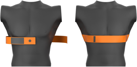
Chest-strap for folding the INYU wearable WBSN in place on the torso. This is applicable for a hand-free use, such as practicing for sports, or overnight screening.
INYU sensor and prototype.
- Front:
- STM32L151RDT6 (ARM Cortex-M3 MCU, 384 kB Flash, 48 kB RAM)
- MPU-6000 (6-axis I²C motion sensor)
- nRF8001 (BLE radio)
- Back:
- AFE4300 (Analog front-end for body impedance measurement)
- ADS1191 (Analog front-end for ECG applications)
Sampling (V-ADC, T-ADC, adaptive knowledge-based sampling)
Action potential of a neuron
Action potential generated by a neuron. An external trigger, called stimulus, will activate the neuron if it is higher than a threshold. In turn, the neuron will transmit the input signal to the following neurons by generating another action potential. For example, in the eye, a photon (the stimulus) will activate a rod cell (a photosensitive neuron) that will transmit the signal to the cortex. Original by Chris 73, updated by Diberri. Modified for this thesis according to the CC-BY-SA 3.0 license.
Adaptive sampling of an ECG
Event-triggered adaptive sampling of an ECG fragment using polygonal approximation. Top: Original signal, sampled at 360 Hz. Bottom: Resulting signal of the adaptive sampling method. The detection of a regular rhythm enables a substantial reduction of the sampling frequency by getting a much coarse representation of the signal. After a rhythm change (second vertical line), the sampling frequency is in- creased to allow capturing the details of the abnormal area. Data source: Record 119 of the MIT-BIH Arrhythmia DB, lead MLII, between 17:10 and 17:24
Uniform V-ADC and level-based T-ADC sampling
Comparison of the working principle of the usual V-ADC and the event-triggered T-ADC.
- As a first step, the micro-controller μC configures the chip responsible for the analog-to-digital conversion, visible as black arrows.
- In the case of both V-ADC and T-ADC, the red lozenges shows the measurement trigger. In the V-ADC case, the trigger happens on regular time intervals, whereas in the T-ADC case, the signal itself triggers the sampling by crossing the configured thresholds.
- The measured value is visible following the red arrows until they reach the axis.
- Whether it is voltage (V-ADC) or time (T-ADC), this analog value needs to be digitized, using a given number of bits, which establishes the resolution of the measure. This conversion is showed with the green arrows. It illustrates the origin of digital noise, making visible the rounding of each sample towards discrete digital values, which are represented as green dots.
- Finally, each sample’s binary value (in green) is returned to the micro-controller μC following the blue arrows.
Wall-Danielsson approximation of the ECG
Wall-Danielsson approximation (black line with round markers •) of the ECG with multiple thresholds, with a maximum allowed error of 1 mV·ms and of 0.4 mV·ms.
Each colored ECG segment (upper part) is split by the algorithm when reaching the maximum error threshold. The corresponding error and the tolerance threshold is depicted in the lower part, with matching colors. Additionally, the dotted line between the plus signs + illustrate a segment being constructed by the algorithm, as its error is below the defined threshold. The red asterisk symbols ∗ are extra samples generated because of the non-optimality of the algorithm. Data source: Record A00848 from the PhysioNet Challenge 2017 database, between t = 2.8s and t = 3.8s
Instantaneous frequencies in the ECG
The ECG, as a solid white line, is superimposed over its spectrogram. Among the three heart-beats in the ECG, the middle one is annotated with each component: the P wave, the QRS complex, and then the T wave. The ECG’s range is from -0.190~mV to +0.296~mV. The evolution of the power for each frequency is illustrated in the background, where high power appear as bright yellow and low power in dark blue. Data source: Record A00848 from the PhysioNet Challenge 2017 database, between t=1.75s and t=4.6s, sampled at 300~Hz. Analysis settings: FFT window: 32 samples, window overlap: 31 samples, FFT size: 1024
Energy breakdown for ECG on Shimmer platform
Energy spent on a WSN for ECG-based cardiac monitoring per second. Three main different strategies are considered for the application:
- Streaming: in this scenario, the signal is sampled and streamed using the platform’s low-power wireless link (IEEE 802.15.4). No signal processing or analysis is performed on the WSN itself.
- Single-lead: in this scenario, a single lead of the ECG is considered. The amplitude and timing information of the nine fiducial points of the beat-beat are extracted, transmitting only the results on the wireless link. While bringing savings energy-wise, the improvement is marginal.
- Two-leads: these two scenarios consider the analysis of the signal using two ECG leads. Using more channels improves the results’ quality concerning the nine fiducial points. As in the single-lead scenario, only the results of the analysis are transmitted wirelessly. The two scenarios only differ by the pre-processing implemented, where the morphological filtering is an optimized approach compared to the cubic spline filtering.
Results from Francisco Rincón, Joaquin Recas, Nadia Khaled, and David Atienza. Development and evaluation of multilead wavelet-based ecg delineation algorithms for embedded wireless sensor nodes. IEEE Transactions on Information Technology in Biomedicine, 15(6):854–863, 2011.
Visual comparison of sensing sample reduction: Compressed Sensing, Uniform Sampling, and Level-Crossing
Reconstruction error between the classical V-ADC, Compressed Sensing, and the event-triggered T-ADC.
All scenarios show a similar number of samples. Due to the low sampling frequency, the V-ADC signal is missing almost completely the R peak from the QRS complex, whereas the T-ADC, relying on an exponential scale of levels, captures the most important feature. The compressed sensing approach has been configured to yield a similar number of samples for the frame considered. Data source: Record A00848 from the PhysioNet Challenge 2017 database, between t=2.8s and t=3.8s
Wireless Sensor Node with T-ADC, V-ADC, Bluetooth, LoRa, and local or remote processing
Structure of the cardiovascular monitoring system, along with the final data sink on a smartphone or in the cloud. Different possibilities of signal acquisition (uniform or event-triggered sampling), data processing (online in the sensor or offline on a remote device) and transmission (short-range Bluetooth Low-Energy (BLE) or mid-range Long Range Wide-area network (LoRa)) are considered. In total, there are eight different scenarios envisioned which can significantly impact the energy budget. The light-dotted arrows are constraints: the ECG processing needs to happen only once, whether it is online or offline.
Algorithms
Support Vector Machine (SVM)
Example of a non-linear SVM.
Decision Tree
Example of a decision tree. It can be also adapted to show the idea of Random Forest with three different base trees.
Neural Network
Example of a neural network with two hidden layers.
Z-filtering
Diagram of a rational transfer function of a filter where a 0 = 1 for normalization. https://ch.mathworks.com/help/matlab/ref/filter.html#buaif7c-5
Obstructive Sleep Apnea
Affected population
Proportion of male adults in the U.S.A. being affected by OSA. In the upper bar, the green part on the right corresponds to people without OSA. The yellow part on the left is divided in two, with a zoom on the lower bar. Among this population of affected people, most of them (the diagonal lines) are undiagnosed. Over the total population considered, only 2.5% are diagnosed with OSA.
OSA classification rate depending on the RR frequency band
Classification accuracy (normalized between 0 and 1) for OSA when varying frequency-band bounds considering the RR-intervals time series. The circular dot is placed at the position of the best normalized frequency band.
From RR-apnea score to ground truth
RR-score for OSA computed from the ECG, along with the chosen threshold. On the lower part of the graph, three representation of Boolean signals:
- from .2 to .3: the direct result of the OSA evaluation, from the apnea RR-score and the threshold,
- from .1 to .2: the unweighted moving average of the direct apnea evaluation, with a window length of 13 minutes,
- from 0 to .1: the OSA labels provided by the database, annotated by an expert.
Data source: Record a19 from the PhysioNet Challenge 2000 ECG-Apnea database
Apnea spectrogram
Spectrogram of the log-power of the RR-intervals series from the recording x32 (PhysioNet Apnea-ECG database) with the labeled OSA logic signal on a same time axis.
Data collection and processing flowchart in the device
Overview of the processing blocks integrated in the device used for the online OSA analysis in the proposed system (INYU). The ECG is first filtered to remove the noise, then the fiducial points are extracted. Finally, the signal is analyzed to detect OSA and cardiac pathologies. Raw ECG data is stored compressed on a memory for further offline analysis by an expert.